
2026 Research Grant Recipients
The ABTA Research Program oversees the competitive, peer-reviewed grant process to ensure funding goes to the most promising brain tumor research. Once awarded, we track each project’s progress and long-term outcomes to measure impact and advance discoveries in the field.
2025 ABTA-Funded Research Projects
The ABTA awarded more than $1.3 million towards 31 grants
The 2025 funded projects span multiple tumor types
- Glioblastoma
- Medulloblastoma
- Metastatic Brain Tumors
- Malignant Glioma
- Diffuse Midline Gliomas
Research areas of focus
- Immunology/Immunotherapy
- Drug Therapies/Experimental Therapeutics
- Epigenetics
- Biomarkers
- Radiation Therapy
- Proteomics
Flexible Research Fund
The ABTA Flexible Research Fund is a flexible approach to target research funding to key gaps in the brain tumor funding landscape. Through these projects the ABTA intends to drive progress in under-recognized areas of research to improve our understanding and the treatment of brain tumors and improve outcomes for patients.

Isabella Glitza, MD, PhD
Institution: The University of Texas MD Anderson Cancer Center
Among solid tumors, metastatic melanoma (MM) has one of the highest risks of spreading to the cerebrospinal fluid (CSF) and lining surrounding the brain, known as leptomeningeal disease (LMD). Patients with LMD face a median overall survival (OS) of only ~4 months. Treatment options are very limited, which must urgently change.
We have shown that delivering Nivolumab (nivo) immunotherapy directly into the CSF (intrathecal injection; IT) can double survival for MM and lung cancer patients with LMD. Further, in melanoma patients without brain or LMD involvement, combining two immunotherapy drugs – nivo and relatlimab (rela) – led to better results than nivo alone. Building on this, we conducted a first-in-human trial testing combined IT/intravenous (IV) nivo/rela in MM patients with LMD.
We also discovered unique gut and oral bacterial patterns (microbiome) linked to clinical outcome in brain metastasis and showed that changing these bacterial patterns affects immune activity in the brain. Our proposal will examine the immune cells in CSF and blood, along with gut and oral microbiome patterns, to assess how they relate to treatment efficacy in our LMD trial. We have successfully enrolled all planned participants and are finalizing sample collection, including blood, CSF, gut and oral microbiome, for our proposed analyses. We are confident that this research will advance our understanding of LMD and could lead to novel treatments for patients facing this devastating complication.
Research Collaboration Grants
The Research Collaboration Grant is a two-year, $200,000 grant, to support interdisciplinary team science projects that combine resources to streamline accelerate progress in the brain tumor field.

Challice Bonifant, MD, PhD and Dean Anthony Lee, MD, PhD (Co-PI)
Institutions:Johns Hopkins University School of Medicine and The Research Institute at Nationwide Children’s Hospital (co-PI institution)

We aim to develop a safer cell therapy for pediatric brain cancers that avoids the toxic effects of chemotherapy, surgery, and radiation on the developing brain. We will engineer natural killer (NK) cells, specialized white blood cells that are the first responders of the immune system, with B7-H3 targeted synthetic receptors, a type of receptor that can localize the NK cells to the tumor. We aim to identify the optimal structure to stimulate NK cell specific anti-tumor attack. We have chosen to target B7-H3 as this protein is highly expressed in lethal pediatric brain tumors. NK cells offer broad innate anti-tumor activity, important for tumors with variable antigen expression. NK cells can also be safely infused from healthy donors to patients without risk of severe toxicity. We plan rapid clinical translation of our developed engineered NK cell therapy and have aligned our evaluation of NK cell activity with the practice on currently open trials, including local injection of NK cells into the tumor site. A key objective for our proposal is comparing clinical NK cell expansion with research methods across the two collaborating sites, including gene insertion and NK cell activation. This proposal addresses major barriers to NK immunotherapy: tumor recognition, sustained activation and clinical grade expansion. By project end we expect an optimized B7-H3 targeted NK cell therapy with demonstrated efficacy and persistence in preclinical models, a validated manufacturing process suitable for multi-center clinical trial, and abundant data generated to support translation to the treatment of high-risk brain tumors.
Basic Research Fellowship
The Basic Research Fellowship is a two-year, $100,000 grant, awarded to post-doctoral fellows who are mentored by established and nationally-recognized experts in the neuro-oncology field.

Kathryn Blethen, PhD
Institution: Duke University School of Medicine
Mentor: Gerald Grant, MD
Tribute: In memory of Keith Shelly
Glioblastoma (GBM) is the most common and aggressive type of adult brain cancer. Glioblastoma is difficult to treat because of two major challenges: 1) the blood vessels in the brain, known as the blood-brain barrier (BBB), are super restrictive and limit the entry of drugs into the tumor, and 2) the immune system is weakened by the tumor, preventing immune cells from killing cancer cells. Focused ultrasound (FUS) is a technology that uses sound waves to temporarily open the BBB to increase drug delivery into the tumor area while also stimulating the immune system. Our data show that FUS increases natural killer (NK) cells, a specific immune cell type that recognizes and kills cancer cells when activated. Although FUS increases the number of NK cells in the brain, it does not activate these cells. Interleukin-15 (IL-15) superagonist, an immune therapy that helps to boost NK cell activity, potently activates NK cells but is too large to cross the BBB. In our study, we will determine if combining FUS BBB opening with IL-15 superagonist increases NK cell activation to further improve survival in mouse models of glioblastoma.
Shuai Wang, PhD
Institution: NYU Grossman School of Medicine
Mentor: Dimitris Placantonakis, MD, PhD
Tribute: TBD
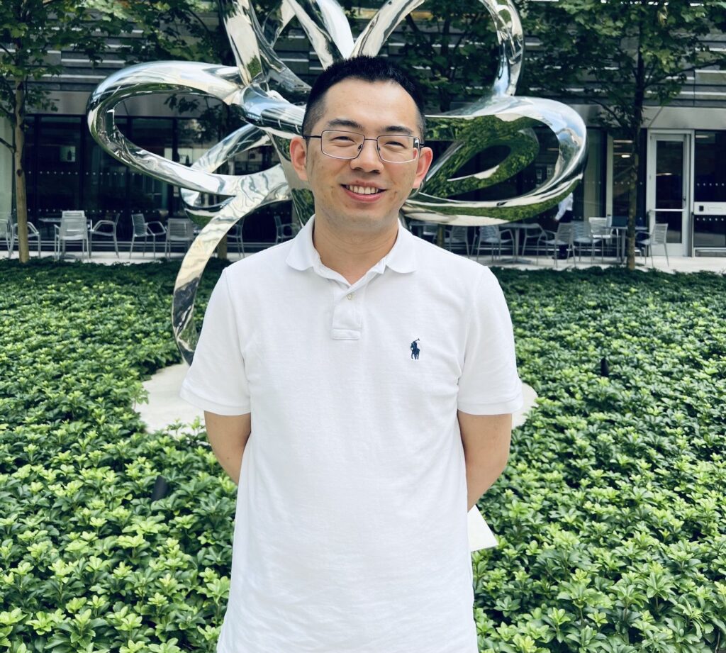
The current treatment of glioblastoma (GBM) has not changed in decades due to lack of new therapies. Recent work indicates that contacts between neurons and tumor cells or interactions among tumor cells generate calcium (Ca2+) signals that promote cancer cell growth. However, how these Ca2+ signals drive tumor growth remains unclear. My work with patient-derived GBM cells and xenografts (a transplanted tissue) has led to the identification of novel underlying mechanisms, which also provide opportunities for therapeutic development. First, I discovered that a major driver of Ca2+ signals is extracellular ATP/ADP, which are known as energy molecules within cells but are released into the extracellular space in tumors. Second, ATP/ADP act through the cell-surface receptor P2RY1, which triggers Ca2+ release into the cytoplasm and nucleus of tumor cells. Third, nuclear calcium activates a protein, CAMK4, which regulates expression of genes that maintain the stemness of tumor cells and drive the aggressive, brain-invasive and immunosuppressive “mesenchymal” state. Finally, I found that pharmacologic inhibition of P2RY1 impairs tumor growth. My proposal aims to a) understand the sources of extracellular ATP/ADP, b) optimize GBM therapy with P2RY1 inhibitors in mouse models, and c) elucidate transcriptional and epigenetic mechanisms downstream of CAMK4 in the nucleus of GBM cells. My research proposal combines novel mechanistic insights with significant translational potential and promises to advance patient care.
Discovery Grants
A Discovery Grant is a one-year, $50,000 grant to support cutting-edge, innovative approaches that have the potential to change current diagnostic or treatment standards of care for either adult or pediatric brain tumors.
Defne Bayik, PhD
Institution: Miller School of Medicine of the University of Miami
Mentor: Antonio Iavarone, MD
Tribute: In memory of Stephanie Lee Kramer
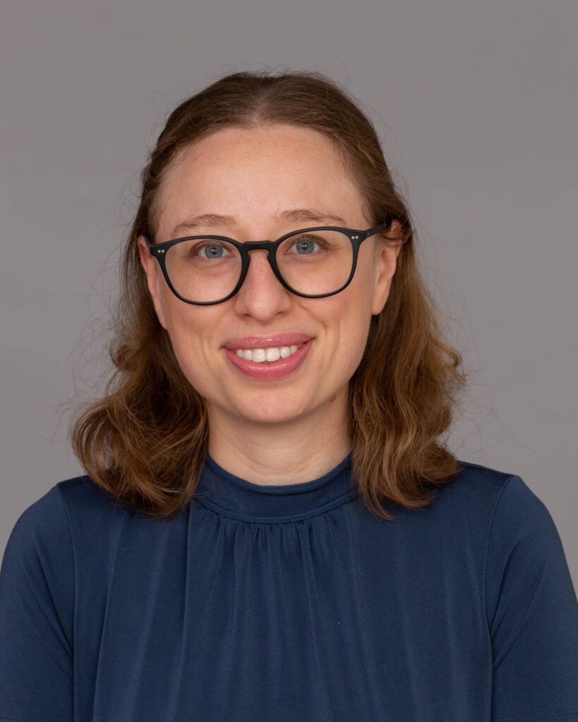
Glioblastoma (GBM) is one of the most common and deadly brain cancers. Unfortunately, treatments that try to directly kill cancer cells have not worked very well. To find better therapies, we need to understand how tumors talk to normal cells in the brain. Astrocytes are the most abundant brain cells, and they normally help keep the brain healthy. However, in GBM, astrocytes can be turned into tumor helpers. Our research has discovered that some GBM cells have a molecule called SLC7A11. SLC7A11 is a transporter that releases glutamate, a chemical messenger the brain cells use to communicate with one another. We discovered that GBM cells release too much glutamate via SLC7A11. This extra glutamate changes how astrocytes behave and may cause them to support the tumor. In this project, we will study how glutamate coming from cancer cells turns astrocytes into tumor-supporting cells. Our goal is to build the foundation for new treatments that prevent GBM cells from hijacking normal brain cells.

Koen Breyne, PhD
Institution: Massachusetts General Hospital
Mentor: Xandra Breakefield, PhD
Tribute: Fully funded by an Anonymous Foundation
Glioblastoma (GBM) is the most aggressive and treatment-resistant brain cancer. Despite surgery, radiation, and chemotherapy, tumors almost always return. One major reason is that GBM creates a powerful “immune shield” that prevents the body’s cancer-fighting cells, called T cells, from working properly. Normally, our immune system can recognize and destroy cancer cells. However, GBM releases high levels of adenosine, a byproduct of cellular metabolism. Adenosine forms part of the tumor’s protective shield by acting as a brake on immune cells. It weakens T cells even before they reach the tumor, limiting the effectiveness of immunotherapies and other treatments. This project aims to release that brake and break the tumor’s immune shield. We will test two innovative strategies. First, we will engineer a therapeutic enzyme that removes adenosine from the tumor environment, restoring immune activity. Second, we will use gene editing to make T cells resistant to adenosine suppression. Together, these approaches are designed to both eliminate the suppressive signal and strengthen the immune response. These strategies will be evaluated using freshly resected human glioblastoma samples to ensure the findings are directly relevant to patients. By restoring the immune system’s ability to fight GBM, this research could lay the groundwork for more effective immunotherapies and new treatment options for patients facing this devastating disease.

Andrea Comba, PhD
Institution: University of Alabama at Birmingham
Mentor: Erwin Van Meir, PhD
Tribute: Fully funded by an Anonymous Foundation
Glioblastoma (GBM) is one of the deadliest brain cancers because current treatments cannot eliminate tumor cells that spread into the surrounding brain. These invasive cells are the main cause of tumor recurrence, yet no therapies effectively target this process. This project focuses on how tumor cells reshape the brain environment to support their spread. Astrocytes are normal brain cells that protect and maintain brain function, but in GBM, they can be reprogrammed to instead help the tumor grow and invade. Our research centers on collagen I, a network of fibers produced by tumor cells that form part of the tumor’s surrounding structure. We believe collagen I acts as a harmful signal that alters astrocytes, turning them from protectors of the brain into active supporters of tumor invasion. We will study how these collagen fibers influence astrocyte behavior and drive tumor spread. This approach represents a fundamental shift in glioblastoma treatment from targeting tumor cells in isolation to disrupting the supportive tumor microenvironment that enables invasion. By preventing astrocytes from being co-opted by tumor cells, this strategy has the potential to limit tumor spread, delay recurrence, and improve patient survival. If successful, this work will establish a new class of microenvironment-targeted therapies and provide a clear path toward clinical translation, addressing one of the most critical unmet needs in brain tumor treatment.

Matthew Gallitto, MD, PhD
Institution: Columbia University
Mentor: David Grosshans, MD, PhD
Tribute: In memory of Brian Bouts
Diffuse midline glioma (DMG) is among the most aggressive childhood brain tumors, with most children surviving only about one year. These tumors grow deep within the brainstem and cannot be surgically removed. Radiation therapy (RT) is the only standard upfront treatment, but recurrence is inevitable. Chemotherapy has largely failed because drugs cannot cross the blood–brain barrier (BBB), a protective lining that blocks most medicines from reaching the brain. Using technology that reads the genetic activity of individual tumor cells, our lab discovered that RT kills only one major cell population within DMG tumors, while a second population survives RT but is sensitive to the drug etoposide. This suggests that combining RT with etoposide could more effectively target the tumor; but some cells still survive both treatments. In this project, we will determine why different tumor cell types respond differently to RT and etoposide, screen hundreds of drugs to find those that kill the remaining resistant cells, and test the most promising combinations in mice using a technique that delivers drugs directly into the brainstem (called convection-enhanced delivery), bypassing the BBB. This technique has already been shown to be safe in children with DMG. These studies will identify rational drug combinations targeting each major tumor cell type and establish a platform to move new therapies toward clinical trials, with the potential to transform treatment for children with DMG.

Ruli Gao, PhD
Institution: Northwestern University
Tribute: Co-funded by BrainUp
Glioblastoma (GBM) is the most common and aggressive brain cancer in adults. Despite surgery, radiation, and chemotherapy, this disease often returns, leading to poor survival. One reason GBM is so difficult to treat is that tumor cells can change and adapt over time. A hidden source of this change comes from how genes produce different versions of proteins through a process called alternative splicing. In cancer, this process can create abnormal protein forms that may help tumors grow and escape treatment. Critically, these abnormal proteins can also generate neoantigens, which are unique signals that the immune system can recognize, thereby triggering the killing of tumor cells that bear these neoantigens. In this project, we will use a cutting-edge technology called long-read single-cell RNA sequencing to study full-length isoforms in individual tumor cells. This allows us to precisely identify tumor-specific protein variants and track how they contribute to neoantigen production and immunogenic responses in both primary and recurrent tumors. By combining advanced computational analysis with protein-level validation, we aim to discover new neoantigens that can be targeted using precise antibody therapies or mRNA vaccines. Our ultimate goal is to develop more effective treatments that prevent GBM recurrence and improve outcomes for patients.

Christopher Garris, PhD
Institution: Massachusetts General Hospital
Mentor: Xandra Breakefield, PhD
Tribute: In honor of Paul Fabbri and all of those who have given on GBM Awareness Day
Glioblastoma (GBM) is an aggressive and often fatal brain cancer, with a median survival of just 15–20 months. Despite decades of research, it remains highly resistant to standard treatments and has not responded to immunotherapies that have improved outcomes in many other cancers. We now know that immunotherapy works best in tumors where immune cells—especially T cells—are already active (“hot” tumors), while tumors lacking these cells (“cold” tumors) typically do not respond. A major reason for this in GBM is its highly suppressive tumor environment. These tumors are packed with immune cells called macrophages, which can make up 30–50% of the tumor and actively shut down immune responses. In contrast, helpful immune cells called dendritic cells—needed to activate T cells—are rare. Previous attempts to overcome this suppression using small-molecule drugs or antibodies have had limited success. Here, we develop a new lipid nanoparticle (mRNA) platform designed to reprogram these immune cells directly within the tumor. These particles deliver instructions that activate key pathways in immune cells, helping turn “cold” tumors into “hot,” immune-responsive ones. We aim to apply this strategy to GBM to test whether reprogramming these cells can overcome immune suppression and make tumors responsive to immunotherapy.

Fada Guan, PhD
Institution: The University of Texas MD Anderson Cancer Center
Mentor: David Grosshans, MD, PhD
Tribute: In memory of Brian Bouts
Glioblastoma is the most aggressive brain tumor, with patients surviving only 12-15 months despite maximum treatment. Doctors cannot safely increase radiation doses because higher doses cause severe, permanent normal brain damage. After decades of research, we have hit a wall—we cannot cure glioblastoma by simply giving more radiation.
This project tests a completely new approach: combining ultra-fast “lightning” radiation with precisely timed electrical pulses to make tumor cells vulnerable while protecting healthy brain. Unlike conventional radiation that takes minutes to deliver, lightning radiation delivers the same dose in milliseconds. The electrical pulses temporarily open tiny pores in tumor cell membranes, allowing radiation damage to accumulate inside cancer cells while normal brain cells remain protected.
If successful, this approach could achieve three breakthroughs: (1) better tumor control with lower total radiation doses, reducing long-term brain damage; (2) condensing six weeks of daily hospital visits into potentially a single treatment session, dramatically reducing burden on patients and families; (3) creating a new treatment strategy for the 12,000 American families facing glioblastoma each year.
Our team combines expertise in radiation physics, brain tumor biology, and electrical pulse technology, with access to one of the few facilities worldwide capable of delivering lightning radiation. Success will lead directly to clinical trials within 3-5 years.

Catalina Lee Chang, PhD
Institution: Northwestern University
Tribute: Co-funded by BrainUp
Chordoma is a rare tumor of the skull base and spine that often returns after surgery and radiation, and there are very few effective drug treatments. Although immune cells are present in many chordoma tumors, they often fail to reach and kill the cancer cells. Our research suggests that this may be caused by fibrous walls inside the tumor, called fibrous septa, which act as barriers. These barriers appear to be built by fibroblasts and enriched in proteins such as collagen and fibronectin that may trap immune cells, especially CD8 T cells, and weaken their activity.
In this project, we will study these barriers using single-cell, spatial, and imaging methods to identify the signals that keep immune cells out of the tumor. We will then test whether breaking down or remodeling these barriers in living slices of patient tumors allows CD8 T cells to move into tumor regions and regain function. The goal of this work is to uncover why the immune system fails in chordoma and to identify new treatment strategies that could make immunotherapy more effective for patients.
Francesca Nazio, PhD
Institution: University of Rome Tor Vergata Institute
Mentor: Grazia Graziani, MD
Tribute: In memory of Stephanie Lee Kramer
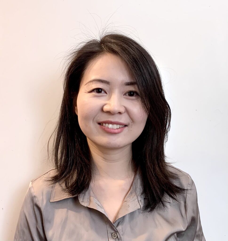
Medulloblastoma is the most common malignant brain tumor in children. Although current treatments—including surgery, radiotherapy and chemotherapy—can be effective for some patients, children with high-risk forms of the disease often experience relapse and poor survival. In addition, radiotherapy can cause serious long-term side effects, especially in very young patients. For these reasons, there is an urgent need to identify new therapeutic strategies that can improve treatment effectiveness while reducing toxicity. Cancer cells survive therapy in part by repairing the DNA damage caused by treatments such as radiation. Tumor cells can activate different DNA repair mechanisms to cope with this stress. One of these mechanisms, called theta-mediated end joining (TMEJ), is controlled by a protein known as POLQ. This repair pathway is more error-prone than normal DNA repair processes but can help cancer cells survive when their genomes become unstable. Our preliminary results suggest that this pathway is particularly active in aggressive forms of medulloblastoma and may contribute to resistance to therapy. In this project, we will investigate how POLQ and the TMEJ repair pathway support tumor growth and resistance to radiation. We will also test whether blocking POLQ can make tumor cells more sensitive to radiotherapy. Understanding this mechanism could reveal a new therapeutic vulnerability in medulloblastoma. In the future, targeting POLQ may help improve treatment outcomes for by enhancing the effectiveness of radiotherapy against aggressive tumors while potentially allowing treatment intensity to be reduced and thereby limiting long-term toxicity.

Christina von Roemeling, PhD
Institution: University of Florida
Mentor: Duane Mitchell, MD, PhD
Tribute: In memory of Stephanie Lee Kramer
Glioblastoma (GBM) and diffuse midline glioma (DMG) are highly aggressive brain tumors with few effective treatments. While immunotherapies such as CAR T cells have shown promise, they are largely ineffective in the brain because T cells cannot efficiently enter or persist within tumors. Even tumors with recognizable targets often lack sufficient T cell infiltration. Although direct delivery into the brain can improve access, it requires invasive surgery, increases cost, and carries risks related to recovery and repeat procedures. We have identified a promising, non-invasive approach to overcome this barrier using low-intensity focused ultrasound (LIFU). LIFU is typically used to temporarily open the blood-brain barrier to improve drug delivery. However, our preliminary data show that LIFU also increases the ability of both natural and engineered T cells to enter brain tumors, suggesting it may actively reshape the tumor environment to support immune cell trafficking. This project will define how LIFU enhances T cell entry and function within brain tumors using advanced imaging and molecular analyses in preclinical models. Understanding this mechanism is essential for safe clinical translation. Ultimately, this work could enable a non-invasive strategy to improve immunotherapy for brain tumor patients while also identifying potential limitations of this approach.
Dionysios Watson, MD, PhD
Institution: Miller School of Medicine of the University of Miami
Mentor: Antonio Iavarone, MD
Tribute: Co-funded by Team Schladt Foundation and an Anonymous Foundation
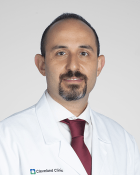
Despite treatment with surgery, radiation, and chemotherapy, most patients with glioblastoma (GBM) survive less than 2 years. Newer therapies that activate the immune system to fight cancer have not worked in GBM. Macrophages and microglia are immune cells that function like orchestra conductors, capable of pushing the immune system to fight against cancer, or shutting it down to help cancer grow. In GBM tumors, macrophages/microglia mostly shut down the immune system’s ability to fight cancer. We propose a new approach to immunotherapy, reprogramming macrophages/microglia to unleash the immune system. We harness the natural ability of these cells to fight bacterial infections. We have isolated microscopic pieces shed by bacteria, called bacterial extracellular vesicles (bEVs), and showed that they activate tumor-promoting macrophages towards more anti-tumor behavior. We showed that bEVs can be safely injected directly into brain tumors or veins of mice, without causing infection or other severe side effects. Treatment with bEVs improved the survival of mice with GBM tumors. In this proposal, we will study how the immune system in GBM tumors of mice changes after bEV treatment. We will also test (in mice) bEVs combined with two other immunotherapies that have been used before in humans. These studies will provide the scientific foundation for future testing of bEVs to overcome the resistance of GBM to immunotherapy, with the ultimate goal increasing survival in GBM patients.

Liangqi (Frank) Xie, PhD
Institution: Cleveland Clinic
Mentor: Jennifer Yu, MD, PhD
Tribute: In honor of Charles “Chip” McKinley Greenlee
Glioblastoma (GBM) is an aggressive and currently incurable brain cancer. Even with surgery, chemotherapy, and radiation, most patients survive only about 15 months after diagnosis. One reason GBM is so difficult to treat is that tumor cells can rapidly adapt and become resistant to therapy. In many GBM patients, a growth-promoting gene called EGFR exists in extra copies outside normal chromosomes. These extra DNA fragments, known as extrachromosomal DNA (ecDNA), are linked to worse patient outcomes. However, we still do not fully understand how they drive tumor behavior or how to target them effectively. In studying tumor cells from 14 GBM patients, we found that about 70% carried EGFR ecDNA. We tested an FDA-approved drug, osimertinib, which blocks EGFR and can enter the brain. Tumors with higher levels of EGFR ecDNA were initially more sensitive to the drug, suggesting a promising treatment strategy. Unfortunately, the cancer cells eventually became resistant. To overcome this resistance, we screened more than 2,100 compounds and discovered that targeting a protein called SMARCA4—an important regulator of DNA packaging—can restore drug sensitivity. Because SMARCA4 functions within several related protein complexes, we will identify which specific complex drives resistance. Pinpointing this target could improve treatment precision while reducing side effects, ultimately leading to safer and more effective therapies for GBM patients.

Eunhee Yi, PhD
Institution: Michigan State University
Mentor: Charles Hong, MD
Tribute: In memory of Stephanie Lee Kramer
Extrachromosomal DNA (ecDNA) is a unique form of circular DNA that resides outside the chromosomes, contains cancer-promoting genes, and is observed in the cancer cells of more than half of glioblastoma patients. Researchers propose that ecDNA significantly contributes to tumor diversity, which can complicate treatment and negatively affect patient outcomes. While many studies have focused on the behavior of ecDNA within individual cells, its movement and function between different cells have not been extensively investigated. Our findings suggest that when a specific cancer cell line from one glioblastoma patient is grown alongside cancer cells from another patient, fragments of ecDNA from the first patient can be detected in the second patient’s cells. This observation raises the possibility that ecDNA is being transferred across different cells. We hypothesize that ecDNA promotes cancer progression by serving as a vehicle for cancer-promoting gene transfer among glioblastoma cells. In this study, we will: 1. Quantify transferred ecDNA and cancer-promoting genes in recipient cells. 2. Determine how the transferred ecDNA affects cancer progression in recipient cells. Through this research, we aim to uncover new behaviors of ecDNA that spread cancer-promoting genes to neighboring cells. This study will also provide valuable insights into the potential for detecting ecDNA in liquid biopsies, which could enable rapid diagnosis of patients with ecDNA-positive glioblastoma.
Jack and Fay Netchin Medical Student Summer Fellowship
A Medical Student Summer Fellowship is a three month, mentor-guided summer research experience, intended to motivate talented medical students to pursue a career in neuro-oncology research.
2025
Lucien Rubinstein Award Recipient

Darwin Kwok, PhD
Institution: University of California, San Francisco
Mentor: Hideho Okada, MD, PhD
Tribute: In Memory of Jameson Taylor, GBM warrior and medical student
Glioblastoma and other brain tumors are very difficult to treat because they almost always come back after standard therapies like radiation and chemotherapy. A major challenge is that these treatments change the tumor in ways that make it harder for the immune system to recognize and attack. Our project set out to understand these changes and to explore how they could be turned into new opportunities for treatment. We discovered that after therapy, brain tumors produce a new landscape of abnormal protein fragments derived from newly formed RNA junctions, which are strands of proteins precursors that connect and change shape. These newly formed RNA junctions are not normally found in healthy cells. These abnormal junctions can create unique protein fragments, or neoantigens, that the immune system could potentially recognize as foreign. By analyzing patient tumors and cell models, we built the first large atlas of these therapy-induced changes. We then used advanced computer tools to predict which of these abnormal fragments could be seen by the immune system and confirmed that many are specific to cancer cells. Even more importantly, we identified a handful of high-priority candidates that appear across multiple patients and treatments, making them especially promising for the development of new immunotherapies such as vaccines or engineered T cells. These findings open up a new way to think about treating brain tumors. By turning the changes caused by therapy into targets for the immune system, we may be able to develop longer-lasting and more effective treatments for patients.

Matthew Abikenari, MSc
Glioblastoma (GBM) is the most aggressive primary tumor of the central nervous system and remains among the deadliest cancers, with a median survival of approximately 15 months despite standard therapy. Although advances in surgery, chemotherapy, and immunotherapy have improved disease management, GBM remains incurable due to profound therapeutic resistance and its ability to suppress antitumor immunity. While tumor-associated macrophages have been extensively studied, the role of tumor-associated neutrophils (TANs) in GBM remains poorly understood. Neutrophils, best known for fighting infections, may play different roles in cancer and could either support or limit tumor growth.
The aim of this study is to determine whether TANs can help fight glioblastoma. Using mouse models, we will examine how neutrophils interact with tumor cells and immune cells, and assess whether the presence of neutrophils is associated with slower tumor growth and disease progression. Understanding whether neutrophils can restrain GBM may reveal new ways to harness the immune system for treatment and improve outcomes for patients with this disease. If successful, this work could change how we think about the innate immune system’s role in brain cancer and open entirely new paths toward therapies for a disease that currently has very few effective options.

Markus Anzaldua-Campos, BA
Institution: Washington University School of Medicine in St. Louis
Mentor: Bhuvic Patel, MD
Tribute: Fully Funded by the Gladiator Project
Glioblastoma (GBM) is one of the most aggressive brain tumors in adults. Even with standard treatment, surgery followed by chemotherapy and radiation, most patients survive less than two years.
GBM is difficult to cure in part because a single tumor contains many different kinds of cancer cells called tumor heterogeneity. Some are more aggressive than others, and when treatment is given, the tumor can shift toward cell types that are better at surviving and resisting therapy. A major challenge in developing new treatments is that many laboratory models used to select drugs for clinical trials have never been exposed to standard chemoradiation, even though most patients receive chemoradiation before entering trials. This mismatch may cause therapies to look promising in the lab but perform poorly in patients.
In this project, we will grow patient-derived glioblastoma organoids, three-dimensional smaller tumors made from a patient’s own tumor tissue and treat them with chemotherapy plus radiation to recreate the post-treatment setting. We will measure how chemoradiation changes the tumor’s cell states and then test whether these changes make the tumor more resistant or sensitive to a panel of drugs. This work will create a more realistic platform for testing therapies after standard treatment, improving how drugs are prioritized for clinical trials and helping guide better strategies for treating glioblastoma.

Noah Drewes, BS
Glioblastoma (GBM) is a deadly brain cancer, and even if patients receive surgery, radiation, and chemotherapy, the tumor comes back in almost all cases. One of the reasons GBM is so hard to treat is because cancer cells learn how to survive even after treatment and can adapt to chemotherapy. If we understand how this process works, we could develop new treatments and help patients with GBM live longer and healthier lives.
One of the most important proteins that leads to cancer survival is HIF-1a, which helps tumor cells grow new blood vessels, invade their surroundings, and behave like stem cells, which in return causes resistance to treatment. In general, HIF-1a is activated by low levels of oxygen, but in our lab, we also found that chemotherapy can cause activation of HIF-1a, even when oxygen levels are normal.
So far, we have found that chemotherapy causes increased HIF-1a in glioblastoma cells. We also found that a protein called CAMK2G can interact with HIF-1a after giving chemotherapy. Ultimately, CAMK2G could help glioblastoma cells survive chemotherapy through activation of HIF-1a.
For this project, we plan to find out if blocking CAMK2G prevents HIF-1a activation, reduces the creation resistant tumor cells, and overall makes glioblastoma less likely to recur after giving chemotherapy. This project could lead to new treatments that make current therapies work better and help prevent glioblastoma from coming back.

Min Jae Kim, BS
Institution: University of Pennsylvania
Mentor: Christina Jackson, MD
Tribute: In Memory of Joe Smeeding
Meningiomas are common brain tumors that often cause symptoms such as weakness, numbness, trouble walking, or confusion. Many meningiomas do not invade the brain, but they can press on and distort the brain’s “wiring” (white matter pathways) that carry signals needed for movement, sensation, balance, and thinking. Today, surgeons mainly estimate a patient’s risks and chances of recovery using tumor size and location on standard scans, along with clinical experience. There is no reliable, patient specific way to determine which pathways are responsible for a person’s symptoms or which pathways must be relieved during surgery to maximize recovery.
This project will use MRI and diffusion MRI, an advanced MRI technique that shows water movement in the brain, to map the white matter pathways that pass through each patient’s tumor and identify symptom specific pathway patterns linked to motor, sensory, gait, and cognitive problems. I will then test whether patients improve when surgery successfully relieves pressure on the pathways most relevant to their symptoms, by comparing pathway patterns related to the surgical cavity and to any remaining tumor after surgery. The expected outcome is a practical imaging based approach that helps predict symptom risk and likelihood of recovery, enabling more personalized counseling and supporting surgical planning that targets the circuits driving symptoms while protecting healthy brain tissue.

Cathy Li, BA
Institution: Cleveland Clinic
Mentor: Justin Lathia, PhD
Tribute: In Honor of Bobby Marshburn
Glioblastoma (GBM) is the most common and aggressive type of brain cancer in adults. Even with surgery, radiation, and chemotherapy, GBM often returns, and survival remains poor. Notably, men are more likely to develop GBM and tend to have worse outcomes than women, but the biological reasons for these differences are not fully understood. One factor that may contribute to these differences is the immune system. A type of immune cell called a regulatory T-cell, or Treg for short, normally helps prevent harmful overactivity of the immune system. However, in GBM, Tregs can protect the tumor by suppressing the immune response that would otherwise help fight cancer. Our early studies show that removing Tregs improves survival to a greater degree in male compared to female mice with GBM, suggesting that Tregs may play a stronger role in suppressing anti-tumor immunity in males. This project will examine why Tregs behave differently in males and females with GBM. First, we will study whether genetic differences between males and females, specifically differences in sex chromosomes, affect Treg levels in brain tumor and peripheral setting. Second, we will test how sex hormones such as estrogen and testosterone influence Treg function and tumor growth. By understanding how genetic and hormonal factors shape immune responses in GBM, this research may help guide the development of more effective, personalized treatments for both male and female patients.

Braxton Morrison, ScM
Institution: University of California San Francisco School of Medicine
Mentor: Jacob Young, MD
Tribute: In Memory of Hong Sun (1958-2025)
Glioblastoma is the most common and most aggressive primary brain cancer in adults. Even with the best available care—surgery, radiation, and chemotherapy—patients often face a poor outlook. Because few new treatments have been approved in the past 20 years, clinical trials are critical for giving patients access to promising therapies and for improving future care. Yet only 8–11% of newly diagnosed glioblastoma patients enroll, in part because identifying appropriate trials is slow and difficult; eligibility rules are lengthy, and the needed details are spread across many pages of medical records.
This project will evaluate whether a “smart assistant” based on large language models (AI tools that can read and organize text) can streamline trial recruitment at UCSF, a tertiary cancer center. We will build an end-to-end system that extracts key clinical details from notes and matches them to trial requirements to generate a personalized list of eligible trials for each patient (Aim 1). We will also create a carefully curated, gold-standard dataset of patients with recurrent glioblastoma to support rigorous validation of future models (Aim 2).
Impact: By reducing missed trial opportunities and staff burden, this work could increase and broaden trial enrollment, improve equity of access to experimental treatments, and accelerate testing of new therapies for glioblastoma.

Saahith Potluri, BS
Institution: Johns Hopkins University School of Medicine
Mentor: Henry Brem, MD
Tribute: Fully Funded by Southeastern Brain Tumor Foundation
Glioblastoma is a devastating brain cancer that is notoriously difficult to treat. One significant obstacle is the “blood-brain barrier,” a protective sheath around the central nervous system that, while vital for health, inadvertently blocks most anticancer medicines from reaching the tumor. This research utilizes a promising genetic therapy using a naturally occurring protein called Growth Arrest-Specific 1 (Gas1). Gas1 acts as a molecular “stop” signal, leading cancer cells to cease dividing and triggering their natural self-destruction. While Gas1 has been shown to significantly shrink glioblastoma tumors, delivering it safely into the brain has remained a major hurdle. Our study proposes a cutting-edge, two-part delivery system: packaging the Gas1 gene into microscopic nanoparticles and using focused ultrasound to gently and temporarily open the brain’s protective barrier at the exact site of the tumor. Focused ultrasound, which is already approved for human use, allows the therapy to be delivered with pinpoint accuracy without the need for invasive surgery. Successful results from this study could potentially provide a roadmap for delivering powerful, tumor-fighting genes directly to the brain. By demonstrating that focused ultrasound can successfully drive Gas1 therapy to diminish GBM in mice, we aim to accelerate its clinical translation. Our goal is to redefine the standard of care and offer new hope to those facing this cancer.

Roland Scott, BA
Institution: Columbia University
Mentor: Brian Gill, MD
Tribute: In Memory of Jameson Taylor, GBM Warrior and Medical Student
Gliomas are aggressive brain tumors that have complex relationships with the surrounding brain tissue. As they grow, they can alter the behavior of nearby brain cells which can lead to seizures. Seizures are common in patients with brain tumors and are often a major source of cognitive and functional impairment at all stages of the disease. Unfortunately, there are no anti-seizure drugs designed specifically for brain tumor patients, and existing drugs work inconsistently. A new drug, AUT00201, holds particular promise as it normalizes the behavior of a select group of neurons which we have previously shown are altered by the presence of tumors. In this study we will test if this drug decreases seizures in a clinically relevant mouse model of glioma. If successful, this could pave the way for future studies in patients with brain tumors who continue to suffer from seizures despite using the current standard of care medications.
Grants Awarded in Collaboration with Other Organizations
Metastatic Brain Tumor Collaborative CNS Metastasis Research Grants
CNS Metastasis Research Grants provide $50,000 over one-year to support researchers to conduct metastatic CNS tumors or leptomeningeal disease-focused projects that are applicable to at least two different primary cancers
Brain Tumor Funders Collaborative
In 2025 the Brain Tumor funders Collaborative (BTFC) awarded two $500,000 grants to support projects focused on Liquid Biopsy for Primary Brain Tumors
